
Electron-Beam Facilities

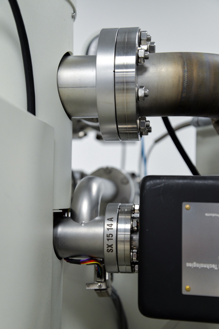
The Department of Earth Sciences is proud to provide analysis using an Electron Microprobe (CAMECA SX-5 FE) and two Scanning Electron Microscopes (FEI Quanta 650 and Thermo Scientific Apreo 2) in its Electron-Beam (E-Beam) facilities. The E-Beam methods are used to study a wide range of natural and artificial solid materials, such as natural volcanic and synthetic glasses, minerals, experimental run products, solar cells, metal alloys and fossiliferous material.
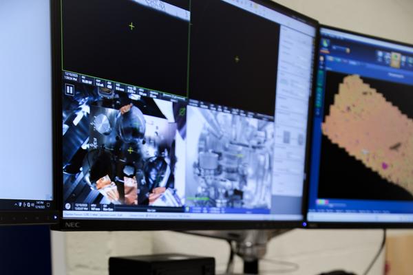
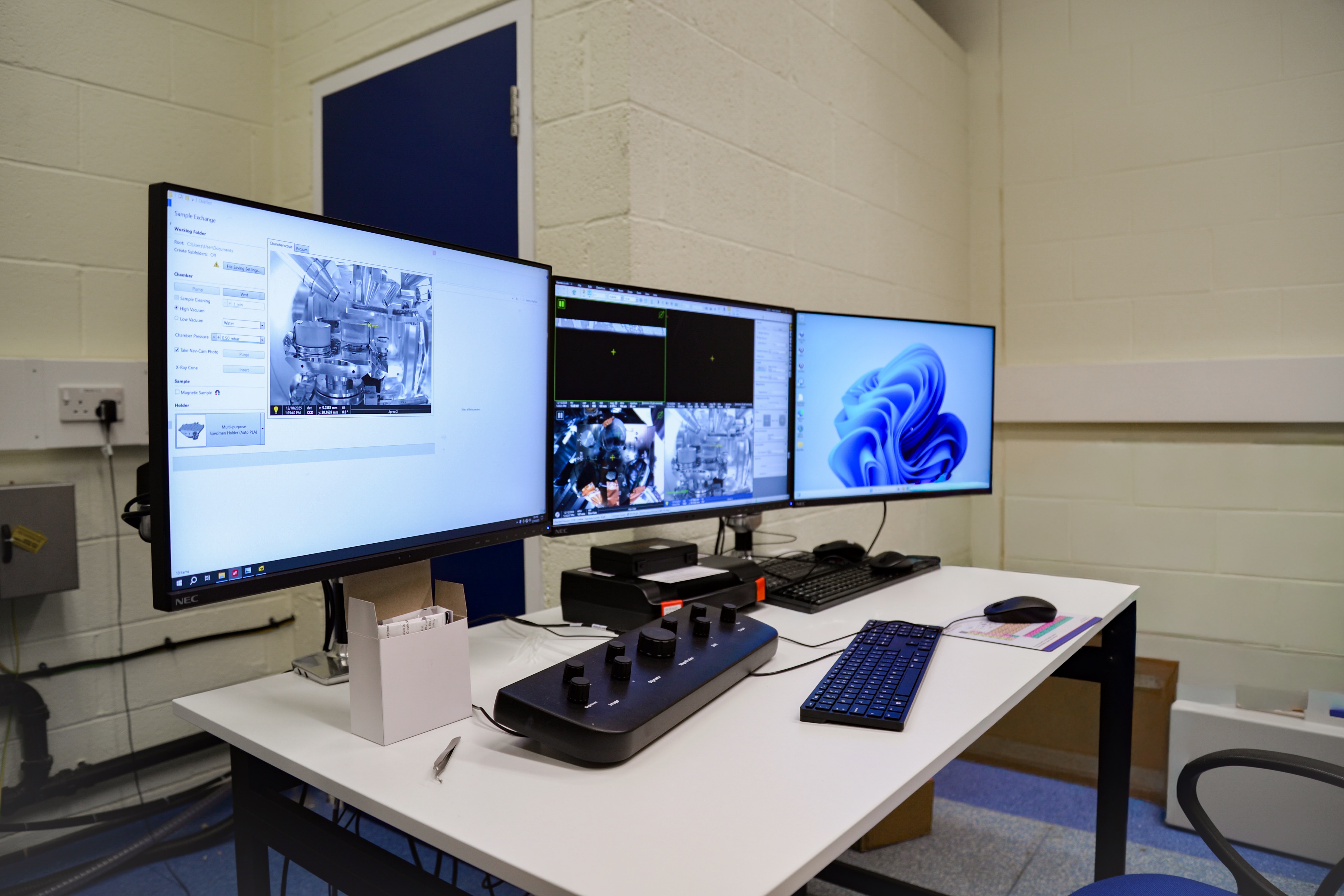
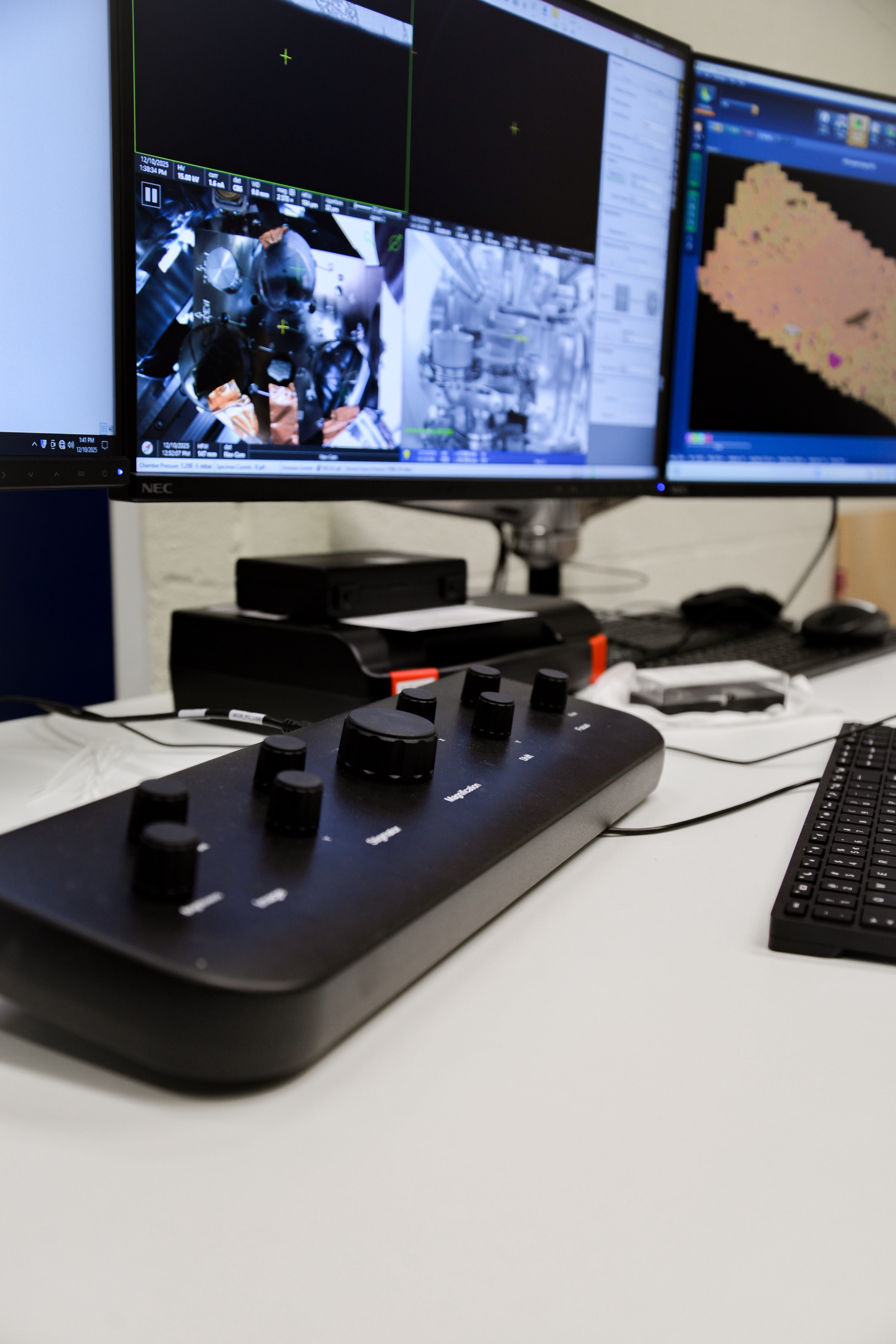
The Scanning Electron Microscope (SEM) is typically employed in imaging surface morphology at sub-mm resolutions and studying major and minor element mapping and quantification using the energy dispersive X-ray spectrometers. The large sample chamber afforded by the SEM is ideal for larger samples (up to 30x30 cm) and its intuitive interface allows for rapid analysis.
The Electron Probe Micro Analyser (EPMA) is capable of high-resolution (~micron), fully quantitative minor and trace-element chemical analysis.
How We Can Support You
We can offer advice at any stage of the research process, including:
- Advice on potential research approaches at the initial stages of inquiry
- Logistics and scheduling
- Aid in sample preparation
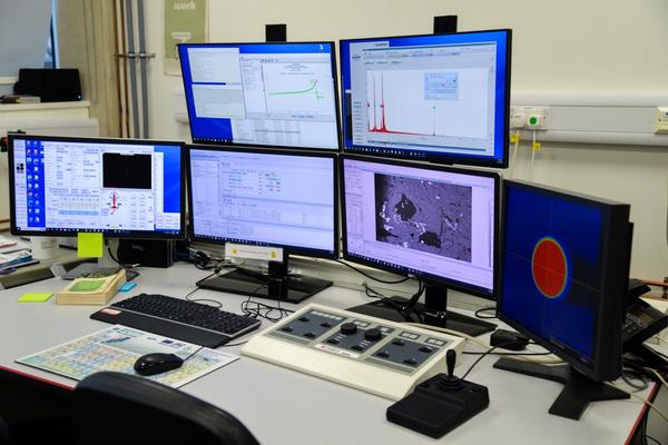
- On-instrument training, calibration and x-ray modelling
- Support when using the equipment and obtaining the data
- Data interpretation and advice on data presentation
The Scanning Electron Microscopes and Electron Microprobe protocols can be precisely tailored to the needs of the researcher. The analytical precision of analyses will depend on the sample itself (composition, sample preparation etc.), the equipment conditions employed, and purpose of research, so please contact us with any questions.
Contact Information
Interested in using the E-Beam facilities? For rates, training, or to schedule use, please contact Andrew Matzen for the Electron Microprobe or Jon Wade for the Scanning Electron Microscopes.
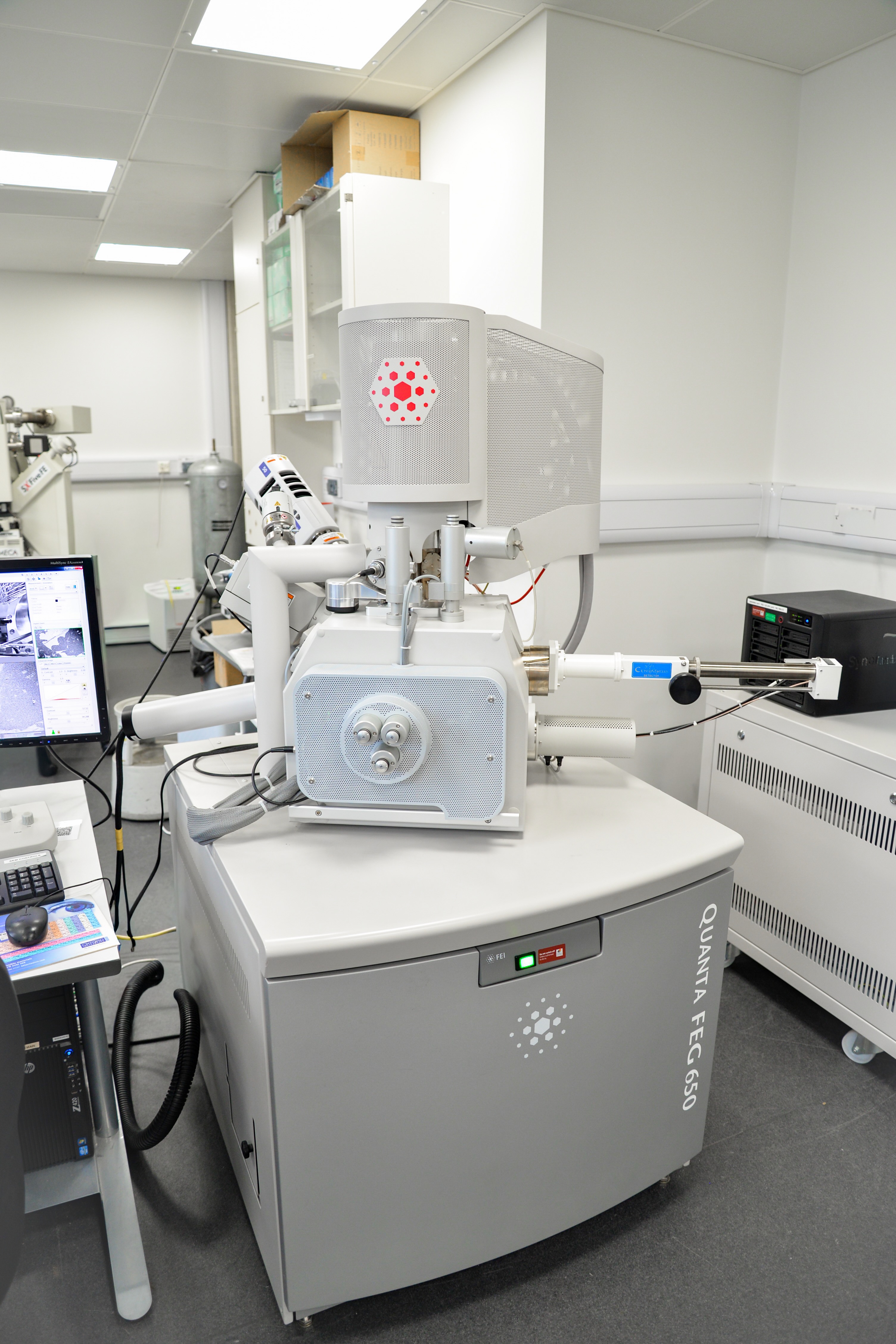
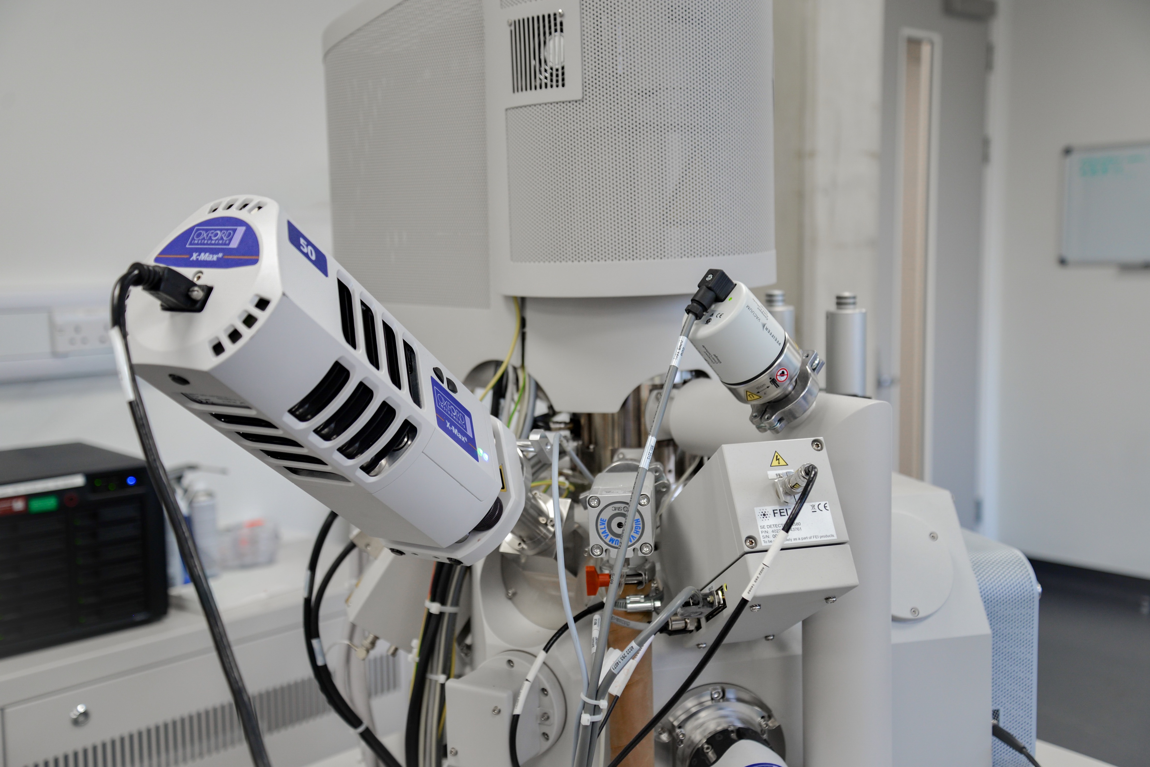
The Earth Sciences Department has access to two Scanning Electron Microscopes (SEMs): a FEI Quanta 650 (installed in 2012) hosted in the Department of Earth Sciences, and a Thermo Scientific Apreo 2 S (installed in 2026) based in the Department of Materials’ David Cockayne Centre for Electron Microscopy. Both instruments are capable of imaging in various modes, as well as the compositional analyses of a range of natural and synthetic materials. Recent uses have included; mineralogical materials, both thin sections and unprepared materials, compositional analyses of high-temperature alloys, biogenic material (lab-grown and natural), extra-terrestrial materials and very high value gemstones.
Key Features

- A field emission electron (FEG) source enabling stable, high-resolution imaging
- Secondary and backscatter electron detectors (enabling both high – and low-vacuum work)
- Oxford Instruments energy-dispersive x-ray spectroscopy (EDS) detector(s) system for rapid major and minor elemental characterization
Approaches and Outputs
- High-resolution (sub-micron) imaging for surface topography (texture and shape) and internal composition
- Secondary electron imaging for high level surface morphology
- Backscattered electron imaging for mean atomic number composition (flat, polished samples) and surface topography
- Energy-dispersive x-ray spectroscopy (EDS) for major and minor (typically >1000 ppm) elemental composition and mapping
- Cathodoluminescence (CL) for electron beam induced light emission arising from certain mineral structures/compositions
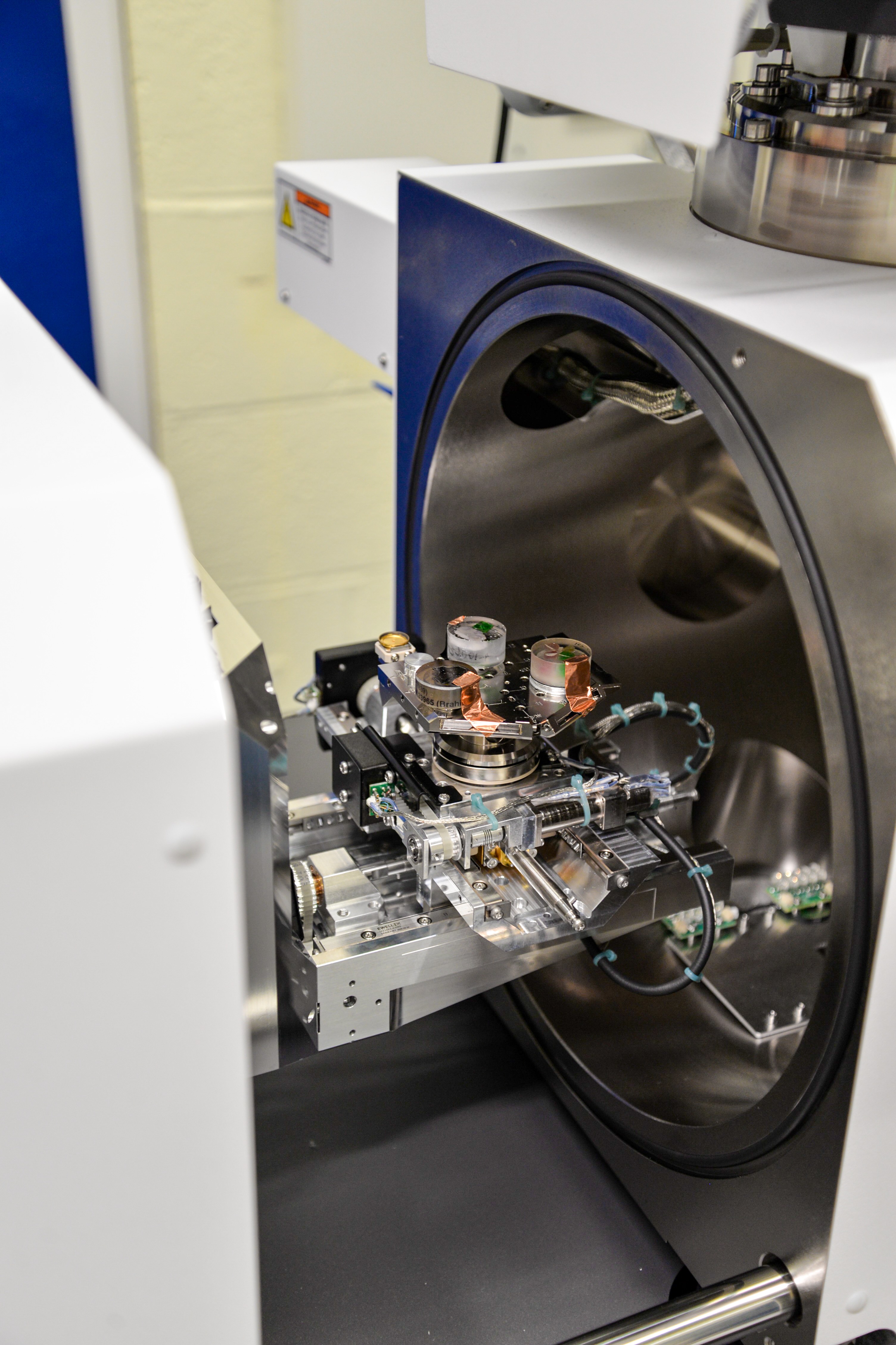
Additional Features and Outputs from the Thermo Scientific Apreo 2 SEM
- Twin 100mm-Oxford Instrument EDS systems for rapid major and minor elemental characterization, mapping and quantification (full standards-based or 'semi-quant')
- In-column hybrid detectors for BSE and SE imaging, delivering much-improved imaging at low accelerating voltages (low kVacc)
- Low vacuum imaging and analyses for uncoated samples
- A retractable BSE detector for highest mean atomic number contrast
- Pseudo-colour CL detector
- In-chamber plasma cleaner for sample surface preparation
- 5-axis stage for large samples (up to ~30x30cm)
Previous Research
-
Compositional analyses of Roman gold coins
- Meteorite and lunar (Apollo) return samples
- High pressure experimental run products
- Fossil material for composition and morphology
- Biological material
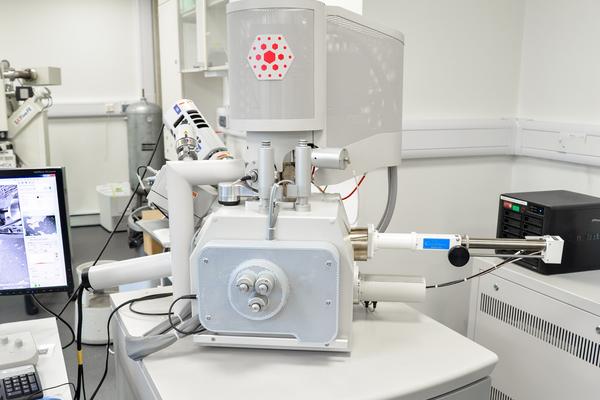
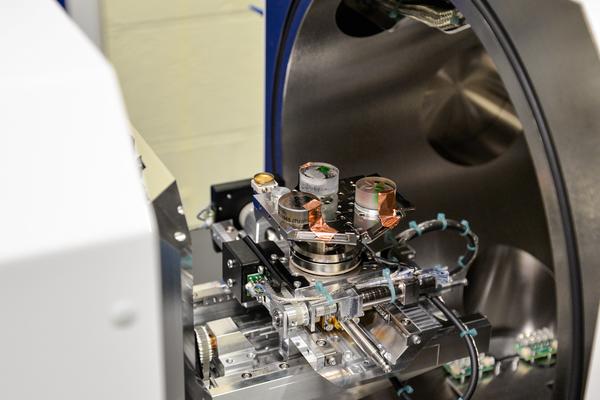


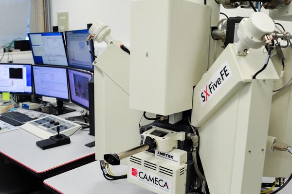
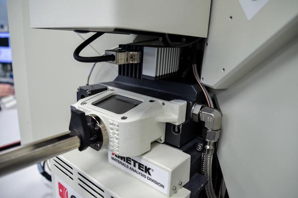
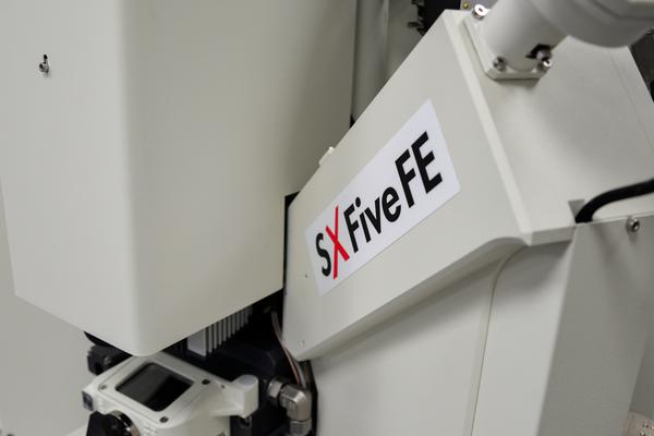
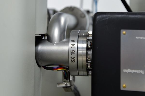
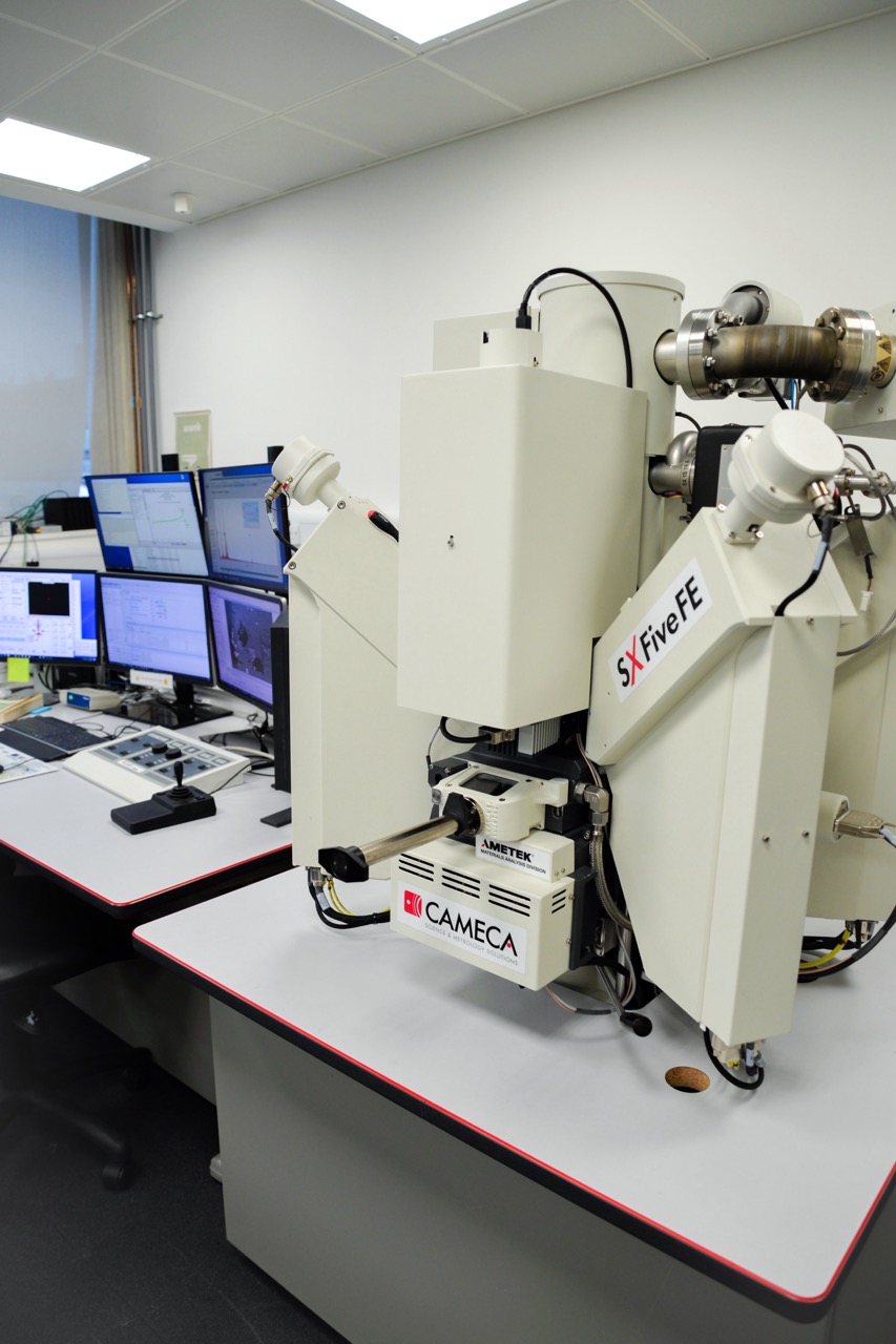
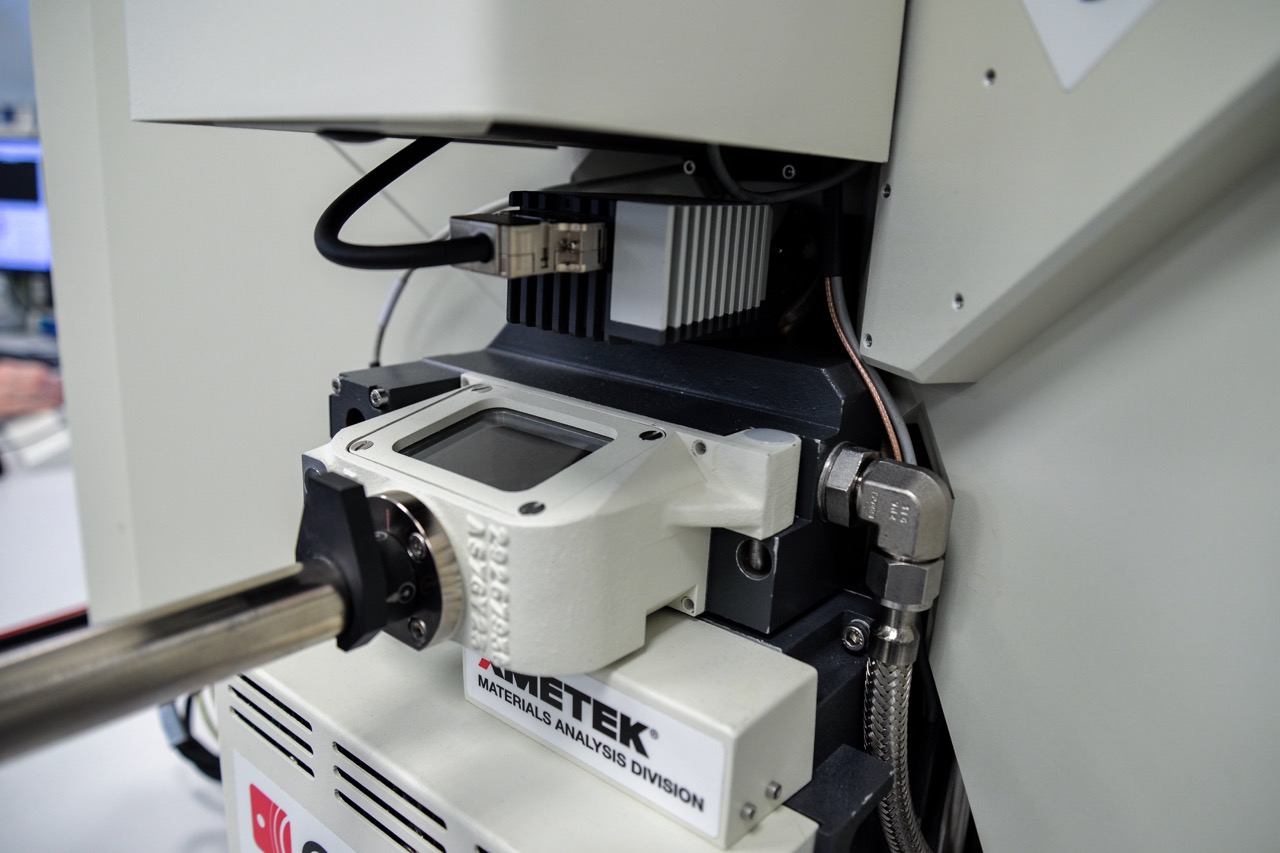
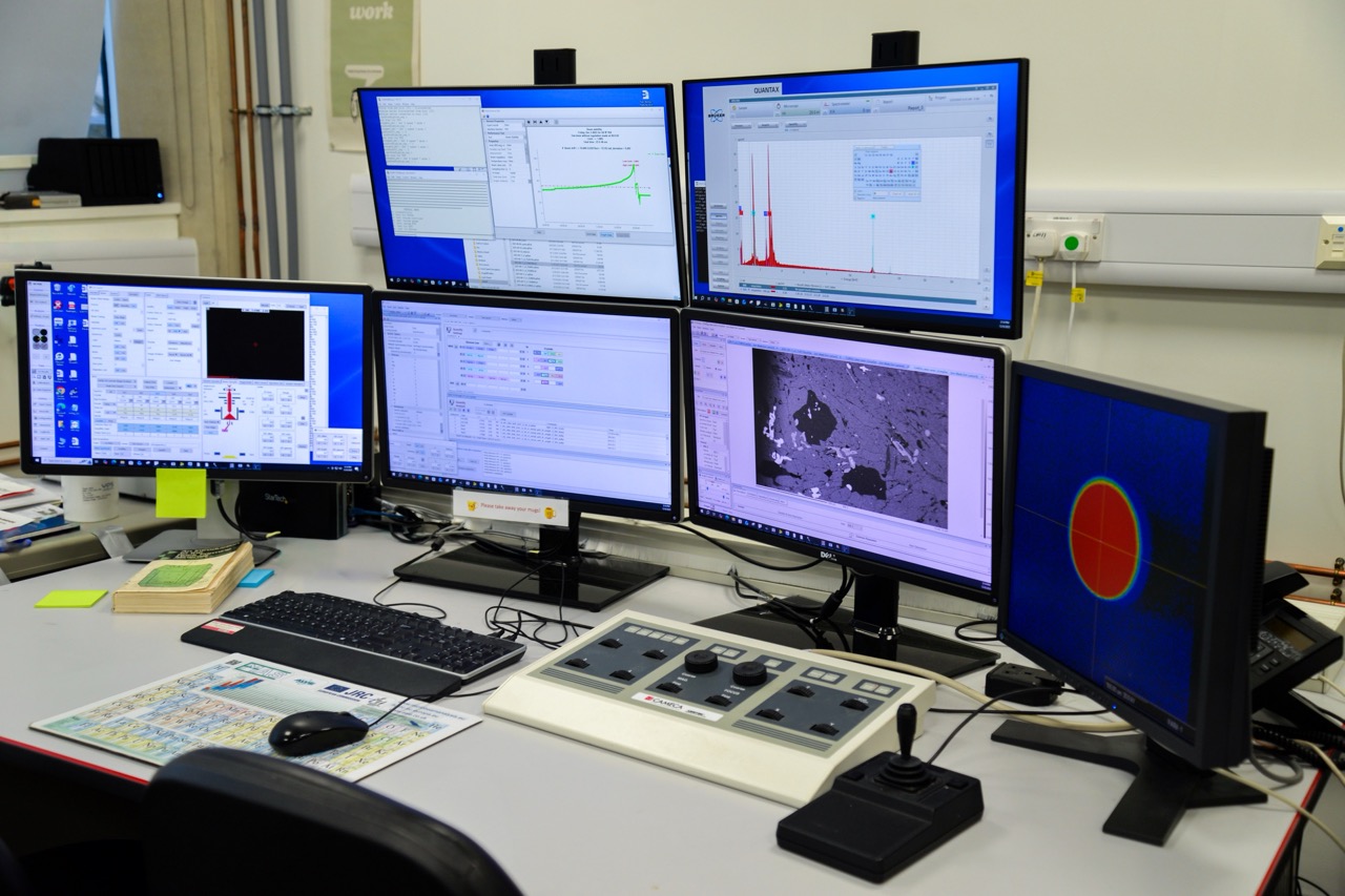
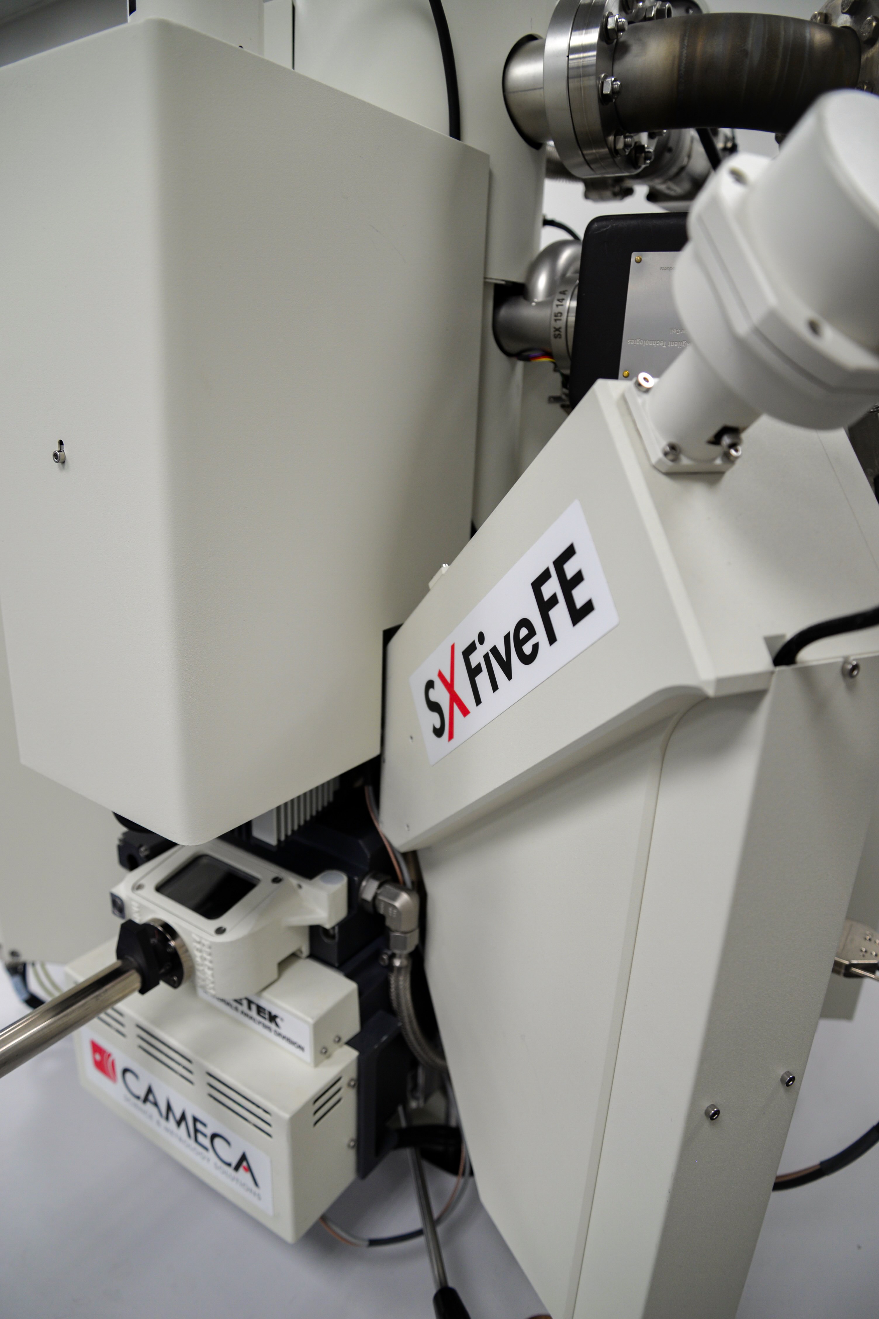
The Earth Sciences Department also houses a CAMECA SX-5 FE, an electron beam micro-analyser (EPMA), commonly referred to as an “e-Probe”, that can quantify chemical compositions at the micron scale with typical detection limits of ~100ppm (0.01 wt%). It can perform major, minor, and trace elemental analysis of elements from carbon to uranium on a variety of solid samples. The e-Probe is a non-destructive approach to chemical analysis, though a flat, polished surface is required. Typically, samples are mounted in vacuum-tolerant epoxy resin or conductive bakelite (metal alloys) and polished prior to analyses.
Key Features
-

A field emission gun provides small spot sizes and sharp images
- Five wavelength-dispersive detectors (WDS) which allow precise analysis at low concentrations
- One energy dispersive detector (EDS)
Approaches and Outputs
- Full quantification: includes full elemental analysis at a single location, or ‘spot’
- Significantly improved energy resolution, and minimised X-ray overlaps when compared to EDS semi-quantification: includes an estimation of elemental composition for approximate concentrations, often used for maps or line profiles
- Secondary electron images which reveal topography
- Backscatter electron images which reveal mean atomic number
Previous Research
- Mineral compositional analyses
- Analyses for thermo-barometry
- Element diffusion profiles in minerals, glasses and metals
- Fully quantified analyses of REE in minerals and transition elements in alloys
- Light element analyses (B, C, N, O and F)


Representatives of Oxford Earth Sciences and Asprey of London
Asprey of London
Oxford Earth Sciences have been collaborating with Asprey of London since 2022 to analyse gem quality jadeite jade (‘jade’). We apply our expertise in mineral analyses to characterise the mineralogy of Asprey’s jade jewellery. As a polycrystalline material, often presented in larger pieces than typical gemmological samples, jade presents some unique analytical challenges. Furthermore, the mechanisms of natural jade formation are debated, and it has not been successfully synthesised in large, gem-sized, masses. This makes gem quality jade both intrinsically rare and geologically interesting. We are delighted to be continuing our work with Asprey over the coming years to develop both our protocols for gem jade analyses and further our understanding of its natural formation mechanisms.